How Many X-Rays Is An MRI Equal To? Unveiling The Radiation Truth
Let’s dive right into the nitty-gritty of medical imaging. If you’ve ever wondered how many x-rays an MRI equals, you’re not alone. This question has been buzzing around healthcare circles for years, and it’s about time we shed some light on it. MRI and x-rays are two of the most common diagnostic tools in the medical world, but they work in completely different ways. While x-rays rely on ionizing radiation, MRIs use magnetic fields and radio waves, which means they don’t expose you to harmful radiation. But just how do they compare? Stick around, and we’ll break it down for you.
When it comes to understanding medical imaging, people often get confused about the risks involved. Terms like radiation, MRI, and x-rays can sound intimidating, especially if you’re not familiar with them. That’s why it’s crucial to know the facts. In this article, we’ll explore the differences between these two imaging techniques and answer the burning question: how many x-rays is an MRI equal to? Spoiler alert: it’s not as straightforward as you might think.
So, whether you’re a curious patient or someone who simply wants to understand the science behind these technologies, you’re in the right place. Let’s demystify the world of medical imaging together and help you make informed decisions about your health. Trust me, this is gonna be good.
- Flix2day Alternative Your Ultimate Guide To Legal Streaming Options
- Flixtor Bz Your Ultimate Streaming Haven
Understanding MRI and X-Ray Basics
What Exactly Is an MRI?
An MRI, or Magnetic Resonance Imaging, is a non-invasive imaging technique that uses powerful magnetic fields and radio waves to produce detailed images of the body’s internal structures. Unlike x-rays, MRIs don’t involve ionizing radiation, which makes them a safer option for many patients. The images produced by an MRI are incredibly detailed, allowing doctors to diagnose a wide range of conditions, from brain injuries to joint problems.
One of the coolest things about MRIs is their ability to provide cross-sectional views of the body. This means doctors can see inside your body layer by layer, giving them a comprehensive understanding of what’s going on. Plus, MRIs are particularly good at capturing soft tissues, like muscles, tendons, and organs, which x-rays can’t always do. So, if you’re dealing with a complex injury or condition, an MRI might be the way to go.
What Makes X-Rays Different?
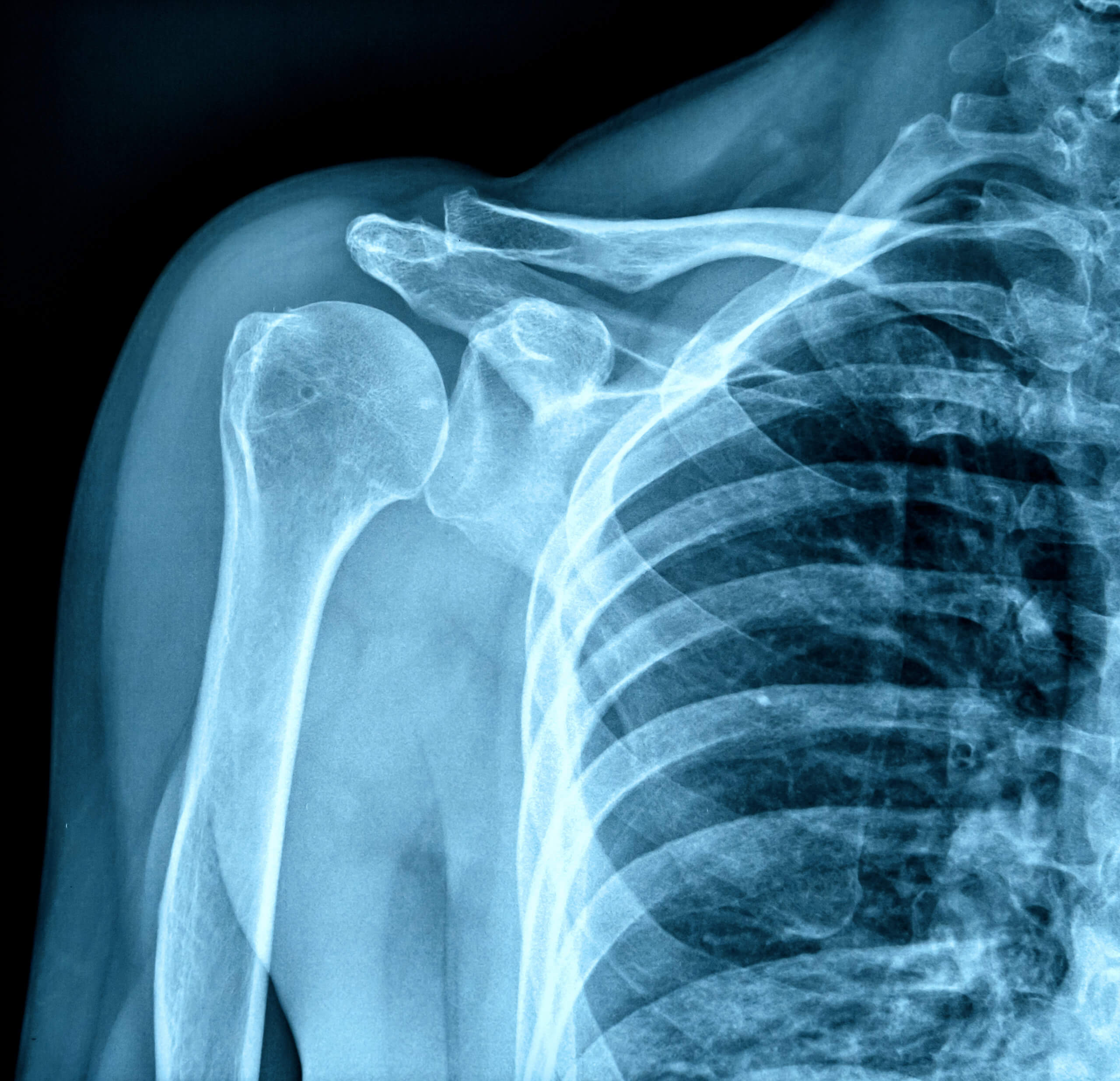
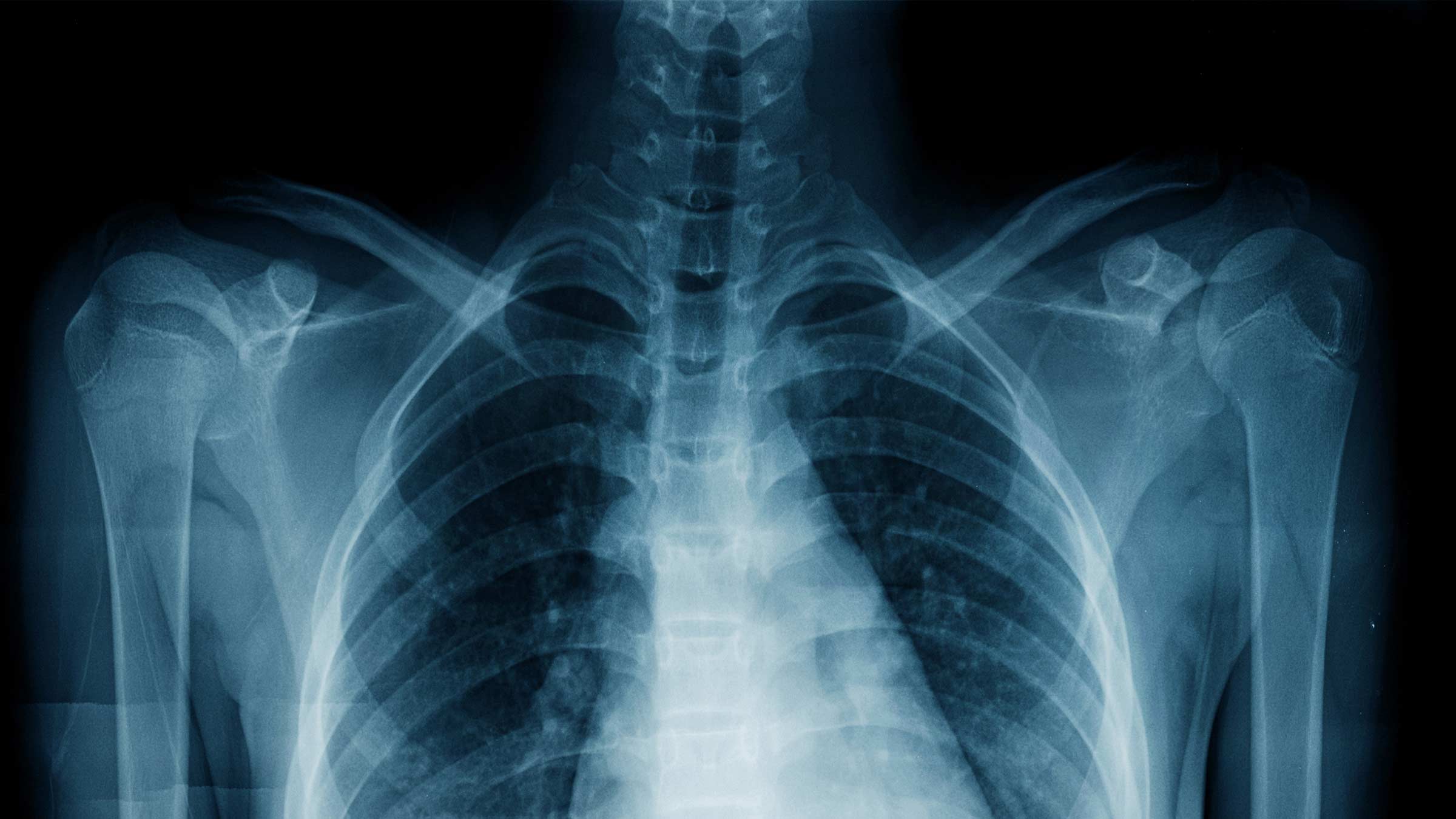
X-rays, on the other hand, use ionizing radiation to create images of the body’s internal structures. They’re most commonly used to diagnose bone fractures, dental issues, and chest problems like pneumonia. While x-rays are quick, efficient, and widely available, they do come with some risks due to their use of radiation. However, the amount of radiation in a single x-ray is generally considered safe for most people.
- Pelisflix2 Your Ultimate Streaming Haven Unveiled
- Streaming Unleashed Your Ultimate Guide To Sflixtvto
Here’s the kicker: the radiation dose in an x-ray is measured in millisieverts (mSv), and the average chest x-ray delivers about 0.1 mSv of radiation. To put that into perspective, we’re exposed to about 2-3 mSv of background radiation just from living on Earth each year. So, while x-rays do involve radiation, the dose is usually pretty low and manageable.
The Radiation Factor: How Many X-Rays Is an MRI Equal To?
Now, let’s tackle the million-dollar question: how many x-rays is an MRI equal to? Here’s the deal—MRIs don’t involve any ionizing radiation at all. That’s right, zero radiation. So, when people ask how many x-rays an MRI equals, the answer is none. MRIs and x-rays operate on completely different principles, so comparing them in terms of radiation exposure doesn’t make much sense.
However, if you’re trying to understand the relative safety of these two imaging techniques, it’s worth noting that MRIs are generally considered safer because they don’t expose you to harmful radiation. That said, MRIs do have their own set of risks, such as claustrophobia for some patients or potential complications for those with metal implants. But when it comes to radiation, MRIs are a clear winner.
Common Misconceptions About Radiation and Imaging
There are a lot of myths floating around about radiation and medical imaging, so let’s clear the air. One common misconception is that all imaging techniques involve radiation. As we’ve already discussed, MRIs don’t use radiation, but CT scans, PET scans, and x-rays do. Another myth is that radiation from medical imaging is always harmful. In reality, the risks depend on the dose and frequency of exposure.
For example, a single x-ray is unlikely to cause harm, but repeated exposure over time could increase your risk of certain health issues. That’s why doctors carefully weigh the benefits and risks before ordering any imaging tests. It’s all about finding the right balance to ensure your safety and get the information needed for an accurate diagnosis.
Breaking Down Radiation Dose Comparisons
Let’s take a closer look at how radiation doses compare across different imaging techniques. As we mentioned earlier, a typical chest x-ray delivers about 0.1 mSv of radiation. A CT scan of the abdomen, on the other hand, can deliver anywhere from 10 to 30 mSv of radiation, depending on the specifics of the test. Now, compare that to an MRI, which delivers exactly 0 mSv of radiation. Zero. Nada. Zilch.
Here’s a quick breakdown of some common imaging techniques and their associated radiation doses:
- Chest X-Ray: 0.1 mSv
- Abdominal CT Scan: 10-30 mSv
- PET Scan: 5-20 mSv
- MRI: 0 mSv
As you can see, MRIs stand out as the safest option when it comes to radiation exposure. But remember, every imaging test has its own set of pros and cons, so it’s important to discuss your options with your doctor.
Why Choose MRI Over X-Ray?
So, why would someone choose an MRI over an x-ray? Well, it all depends on the situation. If you’re dealing with a bone fracture or a chest issue, an x-ray might be the best option. But if you’re dealing with soft tissue injuries, neurological conditions, or joint problems, an MRI is often the way to go. Here are a few reasons why:
- MRIs provide incredibly detailed images of soft tissues, which x-rays can’t always capture.
- MRIs don’t involve ionizing radiation, making them a safer option for repeated imaging.
- MRIs can help diagnose a wide range of conditions, from brain tumors to knee injuries.
Of course, there are some downsides to MRIs as well. They can be more expensive than x-rays, and they might not be suitable for everyone, especially those with metal implants or claustrophobia. But for many patients, the benefits far outweigh the risks.
The Science Behind MRI Technology
How Does MRI Work?
Now that we’ve covered the basics, let’s dive a little deeper into how MRIs actually work. When you undergo an MRI, your body is placed inside a large magnetic field. This magnetic field aligns the protons in your body’s water molecules, and then radio waves are used to disturb those protons. As the protons return to their original alignment, they emit signals that are picked up by the MRI machine and translated into detailed images.
One of the coolest things about MRIs is their ability to produce images in multiple planes. This means doctors can get a 360-degree view of the area being examined, which is incredibly helpful for diagnosing complex conditions. Plus, MRIs can be tailored to focus on specific tissues or structures, giving doctors even more control over the imaging process.
What About Contrast Agents?
Sometimes, doctors use contrast agents during an MRI to enhance the images and make certain structures more visible. These agents are usually injected into the bloodstream and can help highlight blood vessels, tumors, or other abnormalities. While contrast agents are generally safe, some people may experience side effects, so it’s important to discuss any concerns with your doctor beforehand.
When Is MRI the Best Option?
As we’ve already discussed, MRIs are particularly useful for diagnosing soft tissue injuries, neurological conditions, and joint problems. But there are a few specific situations where an MRI might be the best option. For example:
- Diagnosing brain tumors or other neurological conditions
- Assessing joint injuries, such as torn ligaments or cartilage damage
- Evaluating spinal conditions, like herniated discs or spinal stenosis
- Identifying soft tissue abnormalities, such as muscle tears or tendonitis
Of course, every case is different, so it’s important to consult with your doctor to determine the best imaging option for your specific needs.
Addressing Safety Concerns
While MRIs are generally considered safe, there are a few safety concerns to keep in mind. For example, people with certain types of metal implants, like pacemakers or cochlear implants, may not be able to undergo an MRI. Additionally, some patients may feel claustrophobic during the procedure, which can be mitigated by using open MRI machines or providing sedation if necessary.
Another concern is the use of contrast agents, which can cause side effects in some people. While serious reactions are rare, it’s always a good idea to discuss any allergies or medical conditions with your doctor before undergoing an MRI. By addressing these concerns upfront, you can help ensure a safe and successful imaging experience.
What About Pregnant Women?
Pregnant women often have special considerations when it comes to medical imaging. While MRIs are generally considered safe during pregnancy, doctors usually avoid using contrast agents unless absolutely necessary. Additionally, the magnetic fields used in MRIs haven’t been shown to harm developing babies, but it’s always best to err on the side of caution and discuss any concerns with your healthcare provider.
Conclusion: Making Informed Decisions About Medical Imaging
So, there you have it—a comprehensive look at how many x-rays an MRI equals and the differences between these two imaging techniques. To sum it up, MRIs don’t involve any ionizing radiation, making them a safer option for many patients. However, they do come with their own set of risks and considerations, so it’s important to weigh the pros and cons with your doctor.
Remember, the goal of medical imaging is to help diagnose and treat conditions as accurately and safely as possible. By understanding the differences between MRIs and x-rays, you can make more informed decisions about your healthcare. And who knows? You might just impress your doctor with your newfound knowledge of medical imaging technology!
So, what’s next? Leave a comment below and let me know what you think. Are you team MRI or team x-ray? And don’t forget to share this article with your friends and family so they can learn more about the world of medical imaging too. Stay curious, stay safe, and keep asking questions!
Table of Contents
- Understanding MRI and X-Ray Basics
- The Radiation Factor: How Many X-Rays Is an MRI Equal To?
- Common Misconceptions About Radiation and Imaging
- Why Choose MRI Over X-Ray?
- The Science Behind MRI Technology
- When Is MRI the Best Option?
- Addressing Safety Concerns
- What About Pregnant Women?
- Conclusion: Making Informed Decisions About Medical Imaging
- Kormovie Your Ultimate Destination For Korean Movies And Series
- Fzmovieshost Your Ultimate Movie Streaming Destination
Difference Between an MRI and an XRay SI Ortho
X Rays Pictures

The Differences Between X Rays Ct Scans And Mri Scans vrogue.co
