Exploring Extrarenal Pelvis Images: A Deep Dive Into Diagnosis And Understanding
When it comes to extrarenal pelvis images, we’re diving into a world where medical imaging meets diagnostic precision. If you’ve ever wondered about the intricacies of the pelvis and how imaging plays a pivotal role in understanding its conditions, you’re in the right place. This article will unravel the mysteries surrounding extrarenal pelvis images, helping you grasp their significance in modern healthcare.
Let’s face it, medical jargon can sometimes feel like a foreign language. But don’t worry, because we’ll break it down in a way that makes sense. Whether you’re a healthcare professional, a curious individual, or someone seeking clarity about pelvic conditions, this article aims to provide answers and insights.
So, buckle up as we explore the fascinating realm of extrarenal pelvis images. From understanding the basics to uncovering advanced imaging techniques, we’ve got you covered. Let’s get started!
- Pseudoflixpro The Ultimate Streaming Experience You Didnrsquot Know You Needed
- Myflixtor The Ultimate Streaming Destination Youve Been Searching For
What Exactly Are Extrarenal Pelvis Images?
Extrarenal pelvis images refer to visual representations of the pelvic region that focus on structures outside the kidneys. These images are crucial for diagnosing various conditions affecting the bladder, ureters, and surrounding tissues. Think of them as a window into the pelvic cavity, allowing doctors to pinpoint issues with remarkable accuracy.
Now, why are these images so important? Well, the pelvis houses vital organs, and any abnormalities can lead to significant health concerns. By using advanced imaging techniques, healthcare providers can detect problems early, leading to timely interventions and improved patient outcomes.
Why Are Extrarenal Pelvis Images Necessary?
Ever wondered why doctors recommend imaging studies? It’s all about gathering accurate information. Extrarenal pelvis images are essential for diagnosing conditions like urinary tract infections, bladder stones, and even cancer. They help healthcare professionals assess the size, shape, and position of pelvic organs, ensuring nothing is overlooked.
- Solarmovie Pro Your Ultimate Streaming Destination Unveiled
- Myflixerxto Your Ultimate Streaming Destination
Here are some reasons why these images are indispensable:
- Identifying structural abnormalities
- Monitoring treatment progress
- Guiding surgical procedures
- Assessing post-operative changes
By relying on these images, doctors can make informed decisions, ultimately benefiting patients in the long run.
Common Imaging Techniques for the Pelvis
Not all imaging techniques are created equal. When it comes to extrarenal pelvis images, there are several methods at play. Each has its own advantages and is chosen based on the specific condition being evaluated. Let’s take a closer look:
Ultrasound Imaging
Ultrasound uses sound waves to create images of internal structures. It’s non-invasive, safe, and widely used for evaluating the bladder and surrounding tissues. Plus, it’s excellent for detecting fluid accumulation and cysts.
CT Scans
Computed Tomography (CT) scans provide detailed cross-sectional images of the pelvis. They’re particularly useful for identifying tumors, fractures, and other complex conditions. While they involve radiation exposure, the benefits often outweigh the risks in diagnostic scenarios.
MRI Scans
Magnetic Resonance Imaging (MRI) offers unparalleled detail when it comes to soft tissues. It’s ideal for assessing pelvic organs, muscles, and nerves. MRI is especially valuable in cases where precise visualization is critical.
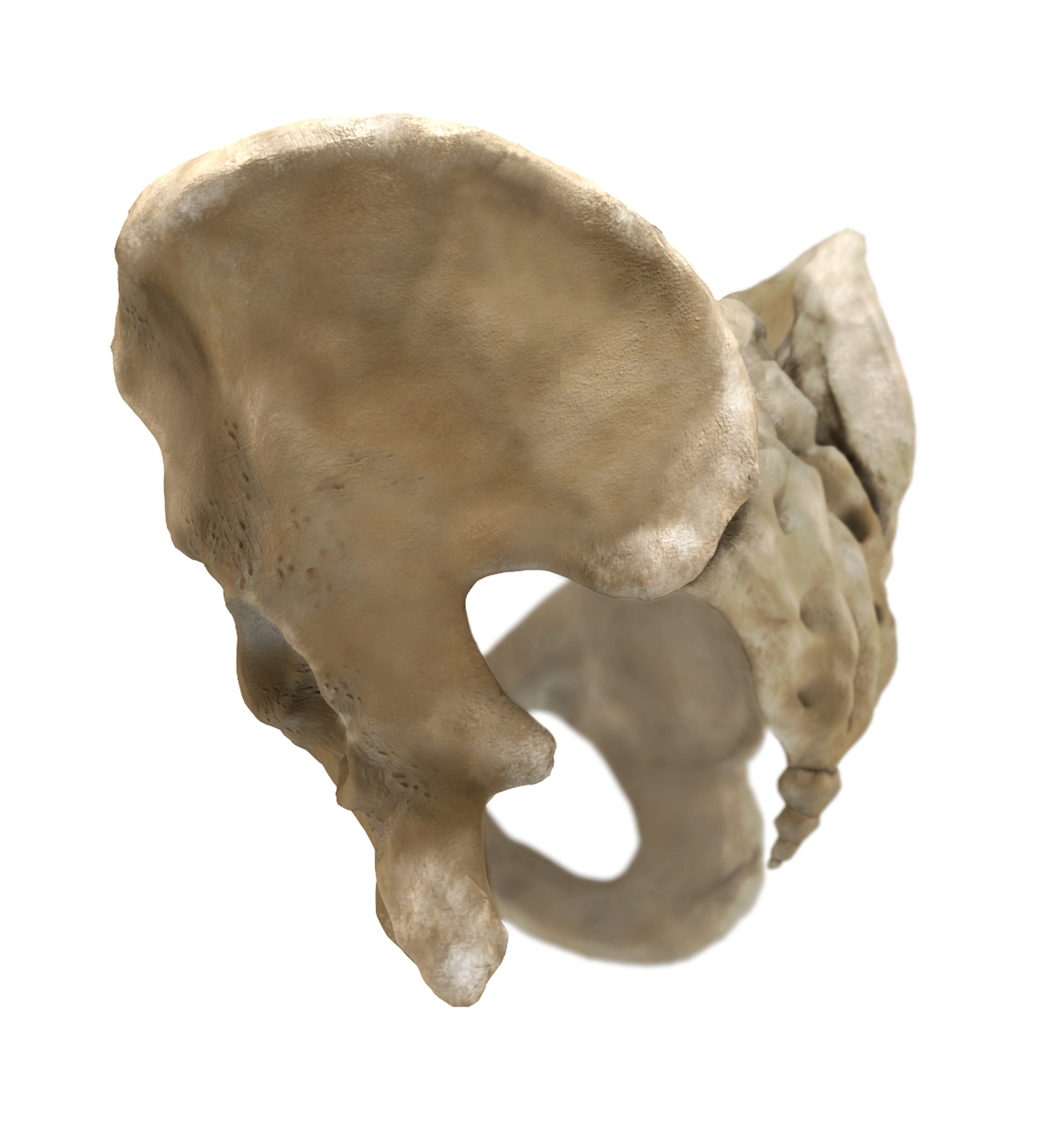
Understanding the Extrarenal Pelvis
The extrarenal pelvis refers to the area surrounding the kidneys, focusing on the bladder, ureters, and adjacent structures. This region plays a vital role in urinary function, making it a key area of interest in medical diagnostics.
Here’s a quick breakdown of what you need to know:
- The bladder stores urine before elimination
- The ureters transport urine from the kidneys to the bladder
- Surrounding tissues and muscles support pelvic organ function
By understanding the anatomy of the extrarenal pelvis, we can appreciate the importance of imaging in maintaining its health.
Applications of Extrarenal Pelvis Images
Now that we’ve covered the basics, let’s dive into the practical applications of extrarenal pelvis images. These images are used in various medical scenarios, each with its own significance:
Diagnosing Urinary Tract Infections (UTIs)
UTIs are a common issue, especially among women. Extrarenal pelvis images help identify inflammation, blockages, and other contributing factors. This ensures accurate diagnosis and effective treatment.
Assessing Bladder Stones
Bladder stones can cause discomfort and complications if left untreated. Imaging techniques like ultrasound and CT scans are instrumental in detecting these stones, allowing for timely intervention.
Monitoring Cancer Progression
In cases of pelvic cancer, regular imaging is crucial for tracking disease progression and treatment effectiveness. Extrarenal pelvis images provide valuable insights, guiding oncologists in their decision-making process.
Challenges in Extrarenal Pelvis Imaging
While imaging technology has advanced significantly, challenges still exist. Factors like patient movement, obesity, and anatomical variations can affect image quality. Additionally, interpreting complex images requires specialized skills and experience.
To overcome these challenges, healthcare professionals rely on cutting-edge technology and continuous education. By staying updated with the latest advancements, they ensure the best possible outcomes for their patients.
How to Prepare for an Extrarenal Pelvis Imaging Procedure
Getting ready for an imaging procedure might seem daunting, but it’s easier than you think. Here are a few tips to help you prepare:
- Follow any fasting instructions provided by your doctor
- Wear comfortable clothing and remove metal objects
- Inform your healthcare provider about any allergies or medical conditions
By taking these steps, you can ensure a smooth and successful imaging experience.
Interpreting Extrarenal Pelvis Images
Once the images are captured, the next step is interpretation. This is where radiologists and other specialists come into play. They analyze the images, looking for abnormalities and patterns that may indicate underlying conditions.
Here’s how the process works:
- Initial review by a trained professional
- Comparison with previous images, if available
- Collaboration with other healthcare providers for a comprehensive diagnosis
Accurate interpretation is key to effective treatment planning, making it a critical component of the diagnostic process.
Benefits of Extrarenal Pelvis Imaging
The benefits of extrarenal pelvis imaging are numerous. From early detection of conditions to guiding treatment decisions, these images play a vital role in modern healthcare. Here are some of the top advantages:
- Non-invasive and safe procedures
- High-resolution images for precise diagnosis
- Reduced need for exploratory surgeries
By leveraging these benefits, healthcare providers can deliver better care and improve patient outcomes.
Future Trends in Extrarenal Pelvis Imaging
As technology continues to evolve, so does the field of medical imaging. Emerging trends like artificial intelligence and 3D imaging are transforming the way we approach extrarenal pelvis imaging. These advancements promise even greater accuracy and efficiency in diagnosing pelvic conditions.
Stay tuned for the latest developments in this exciting field, as they have the potential to revolutionize healthcare as we know it.
Conclusion: Take Action Today
In conclusion, extrarenal pelvis images are a vital tool in diagnosing and understanding pelvic conditions. From identifying infections to monitoring cancer progression, these images provide invaluable insights that guide treatment decisions.
We encourage you to share this article with others who may benefit from the information. If you have any questions or thoughts, feel free to leave a comment below. Together, let’s promote awareness and understanding of extrarenal pelvis imaging.
Table of Contents
- What Exactly Are Extrarenal Pelvis Images?
- Why Are Extrarenal Pelvis Images Necessary?
- Common Imaging Techniques for the Pelvis
- Understanding the Extrarenal Pelvis
- Applications of Extrarenal Pelvis Images
- Challenges in Extrarenal Pelvis Imaging
- How to Prepare for an Extrarenal Pelvis Imaging Procedure
- Interpreting Extrarenal Pelvis Images
- Benefits of Extrarenal Pelvis Imaging
- Future Trends in Extrarenal Pelvis Imaging
- Topmovies Icu Your Ultimate Destination For Movie Buffs
- Why Myflixergs Is Revolutionizing The Streaming Experience

Renal Pelvis

Human Pelvis RoyaltyFree Stock Image 16666090
Human Pelvis A KYU Design
